Imaging myelin degradation in ex vivo prefrontal cortex tissue blocks in Alzheimer’s disease and chronic traumatic encephalopathy
Anna Novoseltseva, Gulce Kureli, Shuaibin Chang, and 10 more authors
Alzheimer’s & Dementia, Aug 2025
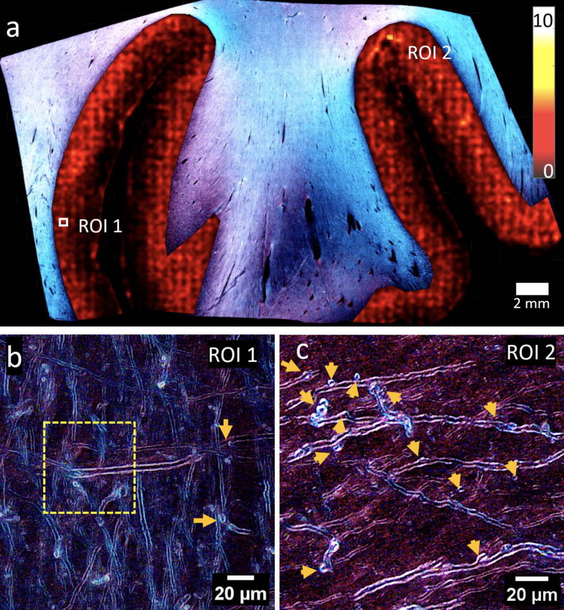
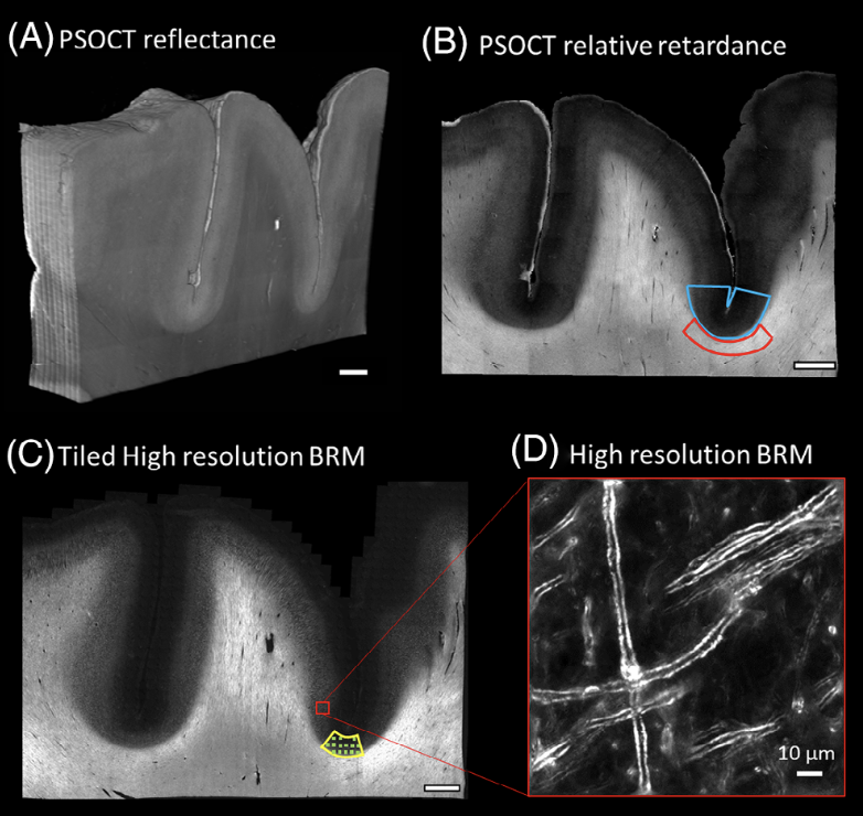
Alzheimer’s disease (AD) and chronic traumatic encephalopathy (CTE) are tauopathies with gray matter (GM) myelin changes that are challenging to assess with standard imaging. New methods are needed to quantify myelin integrity in autopsy brain tissues. We used polarization-sensitive optical coherence tomography (PS-OCT) to measure bulk tissue relative retardance and birefringence microscopy for high-resolution imaging of myelin degradation. Samples included five AD, five CTE, and four age-matched normal controls. When controlling for age and postmortem interval, no statistically significant differences in white matter retardance or GM myelin defect density were observed between groups. The age difference between controls (64 ± 4.7 years, mean ± SD) and disease groups (80.3 ± 7 years) emerged as an important confounding factor. Amyloid beta and tau staining showed weak correlations with myelin defects. Our label-free approach enables large-volume imaging of brain tissue, a valuable tool for studying myelin changes in neurodegenerative diseases. Highlights Multi-modal assessment of myelin integrity using polarization-sensitive optical coherence tomography (PS-OCT) and high-resolution birefringence microscopy. Age emerged as a critical confounding factor; no significant disease differences were found. Weak correlation between myelin defects and deposition of amyloid beta/tau was found in prefrontal gray matter. Label-free optical methods enable high-resolution, large-volume imaging of myelin.